Classifying Hemolytic Anemias: A Multifaceted Approach
By : Dr. Gregory Denomme PhD, FCSMLS (D)
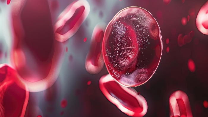
Hemolytic anemias, intricate conditions characterized by the premature destruction of red blood cells (RBCs), can be categorized in two primary ways: based on their root causes and the specific mechanism of hemolysis.
Causes of Hemolytic Anemia
Intrinsic Hemolytic Anemia arises from inherent abnormalities within the red blood cells themselves, which can be either genetic or acquired in nature. These include: Glucose-6-Phosphate Dehydrogenase (G6PD) Deficiency, a genetic disorder that renders RBCs vulnerable and impairs their ability to maintain their structural integrity, leading to their premature destruction; Sickle Cell Disease, hereditary condition characterized by the production of abnormal hemoglobin (HbS), resulting in rigid, sickle-shaped RBCs that are prone to hemolysis and can obstruct blood flow; and Hereditary Spherocytosis, where defects in the proteins that make up the red cell membrane lead to the formation of spherical RBCs which are easily destroyed by the spleen.
Extrinsic Hemolytic Anemia can result from a variety of external factors that directly damage RBCs. These include mechanical causes, such as heart valve prosthetics that can lead to mechanical fragmentation of RBCs; certain infections like malaria, which can cause direct destruction of these vital cells; and Paroxysmal Nocturnal Hemoglobinuria (PNH), An acquired clonal stem cell disorder that increases the sensitivity of RBCs to complement-mediated lysis.
Mechanisms of Hemolysis
Intravascular Hemolysis occurs within the blood vessels and is primarily mediated by the complement system or autoantibodies. This process can be triggered by conditions such as autoimmune hemolysis, where autoantibodies target antigens on the RBC surface, leading to their destruction, or hemolytic transfusion reactions, where transfused blood is incompatible with the recipient's blood type, prompting a rapid immune response.
Key indicators include: Increased plasma free hemoglobin; risk of disseminated intravascular coagulation (DIC); and potential kidney failure due to the difficulty in excreting free hemoglobin.
Extravascular Hemolysis takes place outside the blood vessels, typically in the spleen or liver, where macrophages phagocytize the antibody-coated RBCs. This process is characterized by IgG-coated RBCs being recognized and destroyed by macrophages in these organs.
Key indicators include: Low haptoglobin levels; increased bilirubin; and elevated lactate dehydrogenase (LDH).
Diagnostic indicators
To aid in the diagnosis of hemolytic anemia, healthcare professionals rely on a combination of laboratory findings and clinical observations.
| Hematological Indicators |
|
| Biochemical Markers |
|
| Immunohematological Tests |
|